Scanners in Forensic Medicine
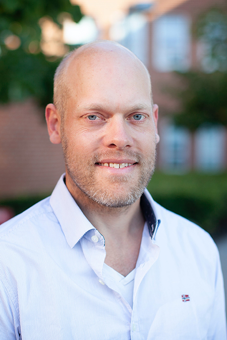
Meet Kasper Hansen
“We plan to devise a method that can simulate a frontal collision in slow motion. Our goal is to collect data and generate knowledge that can provide better traffic-safety measures – it's really exciting.”
Assistant Professor Kasper Hansen, born 1981, is a biologist and has written a PhD on diving physiology. By studying physiological issues in animals, he has acquired unique competences in the use of medical scanning techniques. In recent years, he has specialised in using scanning techniques – primarily CT and MRI – and analyses in new areas within forensic medicine.
Together with researchers from the University of Leicester in England, Kasper Hansen worked on a project that uses deceased people to learn more about what happens in the body during cardiac massage.
"We’ve used a specially-designed research apparatus that compresses the ribcage of cadavers. For each compression step, we do a full CT scan of the body. We subsequently splice the scans together to create a stop-motion film, so we can create a dynamic 4D model of what happens in the body during heart massage,” explains Kasper Hansen. The objective is to enable the researchers to learn more about how to make heart massage as effective and gentle as possible.
Through the use of innovative scanning methods, Kasper Hansen is also working with colleagues from the Department of Forensic Medicine to initiate experimental research in the field of road safety.
"We plan to devise a method that can simulate a frontal collision in slow motion. Our goal is to collect data and generate knowledge that can provide better traffic-safety measures – it's really exciting," he says.

Kasper Hansen is affiliated with the Section for Forensic Imaging and Osteology, but in the summer of 2021 he completed a two-year research stay at the University of Leicester in England, where his research focus was the resuscitation aspect.
Here he has also learned from the success of his British colleagues in CT-angiography, which is used to find narrowing or blockages in the arteries. When the method is used on the living, a contrast agent is introduced in the patient's arteries, and the heart carries the agent around the body. But as the heart is not beating in a deceased subject, a heart-lung machine must be used to recreate a form of circulation. The technique is good in cases where e.g. there is suspicion that the deceased has internal bleeding.
He is also working on developing and refining quantitative and automated scanning analysis methods for post-mortem scans, so these are e.g. able to provide information about the size of internal organs. The method may also result in a new form of support material, for example 3D-printed person-specific models and scanning animations which may be valuable during legal proceedings.
The ability to utilise non-invasive scans to gain knowledge about the cause of death also has an ethical aspect, because in many parts of the world an autopsy is incompatible with religious beliefs. In these areas, the scans can provide valuable insights that would not otherwise be attainable.
"Scanning examinations can also be particularly attractive for examinations of children, because parents can be reluctant to allow an autopsy," explains Kasper Hansen.
The common thread running through all his research projects is the ambition to use non-invasive scanning techniques to acquire knowledge which can provide greater clarity about the cause of death and the body's functions.